English


Introduction
Low back pain is the single largest contributor to years lived with disability worldwide and the most common cause of activity limitation in working-age adults.1,2 Despite intensive research and large healthcare expenditure, outcomes have not improved over recent decades, and the burden of disease continues to grow.1 A central reason is that LBP is, in most patients, not a discrete event but a recurrent condition. Prospective cohort studies and systematic reviews report 12-month recurrence rates ranging from approximately one-third to two-thirds of patients following a first episode, with rates approaching 80% over longer windows.3
This recurrent natural history shifts the central clinical question from “How do we treat the pain?” to “What sub-clinical mechanism persists between episodes, and how can it be reversed?” Across more than three decades of imaging, electromyographic, biomechanical, and interventional research, one mechanism appears repeatedly and is supported by convergent evidence: dysfunction of the lumbar multifidus.9–14 This narrative review summarises the anatomy and biomechanics of the multifidus, the evidence linking its dysfunction to recurrent LBP, the limitations of conventional exercise approaches, and the clinical case for isolated lumbar extension training as implemented in pelvically-restrained device-based rehabilitation.
Anatomy and Biomechanical Role of the Lumbar Multifidus
The lumbar multifidus is the deepest and largest of the paraspinal muscles. Anatomically, it comprises a series of short fascicles arising from the spinous processes and laminae and inserting onto the mamillary processes one to four segments caudal.4,5 Its short lever arm and segmental architecture distinguish it from the long, polysegmental erector spinae and make it ideally suited to controlling motion at individual vertebral levels rather than producing large gross movement.4
Biomechanically, the multifidus contributes the largest single proportion of stiffness to the lumbar segment in vitro,7 and Panjabi’s three-subsystem model of spinal stability positions it as a core component of the active subsystem responsible for maintaining segmental neutral zone control.6 Electromyographic studies have demonstrated that the deep fibres of the multifidus pre-activate in advance of voluntary limb movement, consistent with a primarily anticipatory, stabilising rather than prime-moving role.8 Histologically, the muscle has a high proportion of type I fibres and a dense supply of muscle spindles, both consistent with a postural-stabilising function.
Multifidus Changes in Low Back Pain
Acute, segment-specific atrophy
The first imaging evidence of multifidus involvement in acute LBP was provided by Hides and colleagues at the University of Queensland in 1994.9 Using real-time ultrasound in patients within days of a first acute episode of LBP, the authors demonstrated rapid, unilateral, segment-specific reduction in multifidus cross-sectional area on the side and at the level of symptoms. The atrophy could not be explained by disuse alone, given its acute onset, and was interpreted as a reflex inhibition mediated by nociceptive input.
Failure of spontaneous recovery
The follow-up study by the same group examined whether multifidus atrophy resolved as pain resolved.10 It did not. Patients who had become symptom-free retained the multifidus deficit, demonstrating a dissociation between symptom resolution and tissue recovery. A subsequent randomised trial showed that recovery could be achieved with targeted exercise, and that patients who received such training had significantly lower recurrence rates over the following one to three years than those who received usual care.11 The clinical implication was unequivocal: pain remission is not muscle recovery, and patients who appear to have recovered may continue to carry the dysfunction that produced the original episode.
Chronic structural remodelling
In chronic LBP populations, multifidus changes extend beyond atrophy to include fatty infiltration — replacement of contractile tissue with intramuscular fat — measurable on conventional MRI sequences.12,13 Fatty infiltration of the multifidus is more strongly associated with chronic LBP than fatty infiltration of any other paraspinal muscle and correlates with pain duration, functional disability, and recurrence likelihood.12,13 Translational work in animal models has demonstrated that the response of the multifidus to intervertebral injury is not simple atrophy but a structural remodelling involving adipose and connective tissue deposition driven by local inflammatory signalling.14 The implication is that prolonged dysfunction produces tissue-level changes that may be slower to reverse and more resistant to conventional rehabilitation than acute atrophy alone.
Mechanisms of Dysfunction and Non-Recovery
The persistence of multifidus dysfunction after pain resolution is best understood within the framework of neuromuscular adaptation to pain. Hodges and Tucker’s model of motor adaptation to pain proposes that the nervous system reorganises movement to protect the painful region — typically by reducing activation of deep stabilising muscles and increasing activation of superficial mobilisers.15 This pattern is protective in the acute phase but becomes maladaptive when it persists: the deep stabilisers remain inhibited, the superficial muscles compensate, and the segmental control that the multifidus normally provides is not restored even after pain centralises and resolves.15,16
This adapted motor pattern is not corrected by the resolution of pain itself, nor by the spontaneous return to ordinary activity. Indeed, ordinary daily and recreational activity recruits the multifidus only weakly and almost always in the presence of dominant superficial co-activation,17 providing no specific stimulus for the deep stabiliser to recover. Reversal requires an intervention that selectively loads the multifidus above the inhibitory threshold — which, as the following section argues, conventional exercise rarely achieves.
Why Conventional Exercise Frequently Fails
The recognition that multifidus weakness drives recurrence would, in principle, suggest a straightforward treatment: strengthen the multifidus. In practice, the multifidus is among the most difficult muscles in the body to load directly, and the most commonly prescribed back exercises systematically fail to do so. Three barriers are particularly important:
- Compensation by the global musculature. In any unloaded or free-loaded movement involving the trunk, the superficial erector spinae and the hip extensors — both far stronger than the multifidus — preferentially absorb load. The deep stabiliser is loaded only to the extent that it must contribute to segmental control, which under most conditions is minimal.17
- Hip dominance. Exercises that appear to load the low back — deadlifts, good-mornings, Roman-chair hyperextensions, kettlebell swings — are kinematically hip-hinge movements. The pelvis rotates around the femoral heads while the lumbar spine remains relatively neutral. The lumbar extensors are loaded only to a small fraction of their capacity, and the gluteal and hamstring muscles do the majority of the work.25
- Persistent neuromuscular inhibition. As described above, the adapted motor pattern actively suppresses multifidus recruitment. Voluntary cueing alone is insufficient to overcome this inhibition; the muscle requires repeated, progressive mechanical loading above a recruitment threshold that low-intensity stabilisation work does not reach.22,23
The result is that motor-control and stabilisation exercises produce modest, statistically significant but often clinically marginal improvements in chronic LBP,28,29 and recurrence rates after such programs remain high.3 These programs are not without value, but they do not deliver the intensity required to drive structural adaptation of the multifidus.
Isolated Lumbar Extension Training: The Evidence Base
An alternative paradigm — isolated lumbar extension training with pelvic restraint — has been the subject of sustained clinical investigation since the late 1980s. The original work by Pollock, Graves and colleagues at the University of Florida established two fundamental observations: that lumbar extension strength could be reliably quantified across the full physiological range only when the pelvis was externally stabilised,18,19 and that progressively-loaded isolated lumbar extension produced large gains in lumbar extension strength and corresponding reductions in pain and disability in chronic LBP patients.20
Subsequent trials have replicated and extended these findings across diverse populations and settings. Mayer and colleagues’ functional restoration program demonstrated that quantitative lumbar extension training produced superior return-to-work outcomes compared with conventional rehabilitation in chronic occupational LBP.21 Helmhout et al. compared high-intensity and low-intensity lumbar extension training and found that both produced clinically meaningful improvements in non-specific LBP, supporting the centrality of the modality itself.26 Workplace-based deployment of isolated lumbar extension training reduced low-back injury rates in industrial settings.27
A direct experimental test of the role of pelvic restraint was provided by Smith et al., who demonstrated that lumbar extension training without pelvic stabilisation produced only minimal gains in lumbar extension strength, while the same training with pelvic stabilisation produced large strength gains and significantly greater pain reduction.24 Mechanistic work using electromyography during dynamic extension confirmed that pelvic restraint substantially increases lumbar extensor activation while reducing gluteal contribution.25
The two most comprehensive systematic appraisals of this literature, by Steele and colleagues, concluded that isolated lumbar extension training is more effective than non-isolated alternatives for chronic LBP and provides direct evidence against the broader assumption that lumbar deconditioning cannot be reversed.22,23 Effect sizes for pain and disability outcomes in well-conducted trials of isolated lumbar extension consistently exceed those reported for general exercise, motor-control exercise, and pharmacological therapy.
Clinical Application: Pelvically-Restrained Device Technology
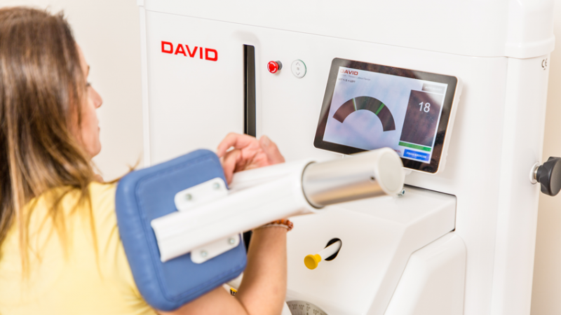
The principles established by the literature above — pelvic restraint, full range-of-motion loading, progressive resistance, and quantification — define the requirements for effective lumbar extensor rehabilitation. The DAVID G110 Lumbar Extension device was engineered specifically to operationalise these requirements in a routine clinical workflow.
Three design features are central:
- Multi-point pelvic and femoral restraint. The pelvis is locked between footplate, thigh restraint, and ischial counterforce before movement begins. With the hips effectively excluded from the kinetic chain, the lumbar extensors — including the multifidus at each segment — become the prime movers. The compensation pattern that defeats free-loaded back exercise is eliminated at the level of the equipment.
- Quantitative assessment integrated with training. The G110 measures isometric and dynamic lumbar extension strength through the full physiological range of motion, generating an objective strength curve. This permits identification of range-specific deficits, comparison against normative reference data, and longitudinal tracking of recovery independently of subjective pain reports — which characteristically lag tissue change.
- Calibrated progressive loading. Resistance adjusts in small, repeatable increments matched to the individual’s measured capacity. Each training session can target a specific portion of the strength curve and deliver a stimulus above the adaptive threshold, while avoiding the under-loading that characterises most rehabilitation programs and the over-loading that risks symptom provocation.
The clinical workflow these features support is straightforward: assess, identify deficit, prescribe individualised progressive loading, reassess. The model treats multifidus dysfunction as a measurable, modifiable, and re-measurable biological phenomenon — not as a clinical inference from pain behaviour alone.
Discussion
The evidence reviewed here supports a coherent and clinically actionable model of recurrent LBP. In most patients, recurrence is not a series of unrelated episodes but the expression of a persistent, sub-clinical mechanism: a segmentally weakened and structurally remodelled multifidus that allows the lumbar spine to be loaded with insufficient segmental control, repeatedly. Pain marks the moments at which load exceeds capacity. Capacity, between episodes, does not spontaneously return to normal — and is not restored by general exercise, motor-control training of marginal intensity, or pharmacological management.
Recovery of the multifidus requires what the broader resistance training literature would predict for any other inhibited muscle: selective loading above the adaptive threshold, applied progressively over weeks to months, with objective measurement to drive prescription. The principal barrier to delivering this in standard clinical practice is the mechanical impossibility of isolating the lumbar extensors without pelvic restraint. Device-based pelvic stabilisation removes that barrier and reproducibly delivers the loading conditions under which the multifidus and the other lumbar extensors adapt.
This framing has implications for both clinical reasoning and care pathway design. Diagnostically, it argues for the inclusion of objective lumbar extension strength testing in the assessment of any patient with recurrent or chronic LBP. Therapeutically, it positions isolated, progressively-loaded lumbar extension not as one of many equivalent exercise modalities but as the modality most directly addressing the underlying mechanism of recurrence. Public health and occupational settings — where Foster and colleagues have called for more mechanism-specific and less generic LBP interventions — may particularly benefit from the predictability and measurability of this approach.30
Several limitations of the available evidence should be acknowledged. Trials of isolated lumbar extension have varied in sample size, comparator, and training protocol, and direct head-to-head trials against contemporary motor-control or graded-activity programs remain relatively few. Imaging-based evidence for multifidus recovery with specific lumbar extension training, though present, is less abundant than the symptomatic and strength evidence. The animal models that have most clearly demonstrated structural remodelling of the multifidus are not perfectly translatable to human clinical practice. These gaps argue not against the framework but for further work — including imaging-based outcome studies and pragmatic comparative effectiveness trials embedded in routine clinical practice.
Conclusion
Recurrent low back pain is, for most patients, the clinical surface of a persistent mechanical-biological problem: dysfunction of the lumbar multifidus that does not resolve spontaneously and is not addressed by the most commonly delivered exercise interventions. The principles of effective rehabilitation — pelvic restraint, progressive loading, and objective measurement — have been established by more than three decades of consistent evidence. Device-based isolated lumbar extension implements these principles in clinical practice. Treating the alarm is necessary; treating the system is what prevents the next episode.
References
- Hartvigsen J, Hancock MJ, Kongsted A, et al. What low back pain is and why we need to pay attention. Lancet. 2018;391(10137):2356–2367.
- GBD 2021 Low Back Pain Collaborators. Global, regional, and national burden of low back pain, 1990–2020, its attributable risk factors, and projections to 2050: a systematic analysis of the Global Burden of Disease Study 2021. Lancet Rheumatol. 2023;5(6):e316–e329.
- da Silva T, Mills K, Brown BT, Herbert RD, Maher CG, Hancock MJ. Risk of recurrence of low back pain: a systematic review. J Orthop Sports Phys Ther. 2017;47(5):305–313.
- Macintosh JE, Bogduk N. The biomechanics of the lumbar multifidus. Clin Biomech. 1986;1(4):205–213.
- Macintosh JE, Valencia F, Bogduk N, Munro RR. The morphology of the human lumbar multifidus. Clin Biomech. 1986;1(4):196–204.
- Panjabi MM. The stabilizing system of the spine. Part I. Function, dysfunction, adaptation, and enhancement. J Spinal Disord. 1992;5(4):383–389.
- Wilke HJ, Wolf S, Claes LE, Arand M, Wiesend A. Stability increase of the lumbar spine with different muscle groups. A biomechanical in vitro study. Spine. 1995;20(2):192–198.
- Moseley GL, Hodges PW, Gandevia SC. Deep and superficial fibers of the lumbar multifidus muscle are differentially active during voluntary arm movements. Spine. 2002;27(2):E29–E36.
- Hides JA, Stokes MJ, Saide M, Jull GA, Cooper DH. Evidence of lumbar multifidus muscle wasting ipsilateral to symptoms in patients with acute/subacute low back pain. Spine. 1994;19(2):165–172.
- Hides JA, Richardson CA, Jull GA. Multifidus muscle recovery is not automatic after resolution of acute, first-episode low back pain. Spine. 1996;21(23):2763–2769.
- Hides JA, Jull GA, Richardson CA. Long-term effects of specific stabilizing exercises for first-episode low back pain. Spine. 2001;26(11):E243–E248.
- Kjaer P, Bendix T, Sorensen JS, Korsholm L, Leboeuf-Yde C. Are MRI-defined fat infiltrations in the multifidus muscles associated with low back pain? BMC Med. 2007;5:2.
- Goubert D, Oosterwijck JV, Meeus M, Danneels L. Structural changes of lumbar muscles in non-specific low back pain: a systematic review. Pain Physician. 2016;19(7):E985–E1000.
- Hodges PW, James G, Blomster L, et al. Multifidus muscle changes after back injury are characterized by structural remodeling of muscle, adipose and connective tissue, but not muscle atrophy: molecular and morphological evidence. Spine. 2015;40(14):1057–1071.
- Hodges PW, Tucker K. Moving differently in pain: a new theory to explain the adaptation to pain. Pain. 2011;152(3 Suppl):S90–S98.
- Hodges PW, Richardson CA. Inefficient muscular stabilization of the lumbar spine associated with low back pain. A motor control evaluation of transversus abdominis. Spine. 1996;21(22):2640–2650.
- MacDonald DA, Moseley GL, Hodges PW. The lumbar multifidus: does the evidence support clinical beliefs? Man Ther. 2006;11(4):254–263.
- Pollock ML, Leggett SH, Graves JE, Jones A, Fulton M, Cirulli J. Effect of resistance training on lumbar extension strength. Am J Sports Med. 1989;17(5):624–629.
- Graves JE, Pollock ML, Carpenter DM, et al. Quantitative assessment of full range-of-motion isometric lumbar extension strength. Spine. 1990;15(4):289–294.
- Risch SV, Norvell NK, Pollock ML, et al. Lumbar strengthening in chronic low back pain patients. Physiologic and psychological benefits. Spine. 1993;18(2):232–238.
- Mayer TG, Gatchel RJ, Mayer H, Kishino ND, Keeley J, Mooney V. A prospective two-year study of functional restoration in industrial low back injury. An objective assessment procedure. JAMA. 1987;258(13):1763–1767.
- Steele J, Bruce-Low S, Smith D. A review of the clinical value of isolated lumbar extension resistance training for chronic low back pain. PM R. 2015;7(2):169–187.
- Steele J, Bruce-Low S, Smith D. A reappraisal of the deconditioning hypothesis in low back pain: review of evidence from a triumvirate of research methods on specific lumbar extensor deconditioning. Curr Med Res Opin. 2014;30(5):865–911.
- Smith D, Bissell G, Bruce-Low S, Wakefield C. The effect of lumbar extension training with and without pelvic stabilization on lumbar strength and low back pain. J Back Musculoskelet Rehabil. 2011;24(4):241–249.
- San Juan JG, Yaggie JA, Levy SS, Mooney V, Udermann BE, Mayer JM. Effects of pelvic stabilization on lumbar muscle activity during dynamic exercise. J Strength Cond Res. 2005;19(4):903–907.
- Helmhout PH, Harts CC, Staal JB, Candel MJ, de Bie RA. Comparison of a high-intensity and a low-intensity lumbar extensor training program as minimal intervention treatment in low back pain: a randomized trial. Eur Spine J. 2004;13(6):537–547.
- Mooney V, Kron M, Rummerfield P, Holmes B. The effect of workplace based strengthening on low back injury rates: a case study in the strip mining industry. J Occup Rehabil. 1995;5(3):157–167.
- Searle A, Spink M, Ho A, Chuter V. Exercise interventions for the treatment of chronic low back pain: a systematic review and meta-analysis of randomised controlled trials. Clin Rehabil. 2015;29(12):1155–1167.
- Saragiotto BT, Maher CG, Yamato TP, et al. Motor control exercise for chronic non-specific low-back pain. Cochrane Database Syst Rev. 2016;(1):CD012004.
- Foster NE, Anema JR, Cherkin D, et al. Prevention and treatment of low back pain: evidence, challenges, and promising directions. Lancet. 2018;391(10137):2368–2383.